

Abstract
Infection of phyllosphere (stems, leaves, husks, and grains) by pathogenic fungi reduces the wheat yield and grain quality. Detection of the main wheat pathogenic fungi provides information about species composition and allows effective and targeted plant treatment. Since conventional procedures for the detection of these organisms are unreliable and time consuming, diagnostic DNA-based methods are required. Nucleic acid amplification technologies are independent of the morphological and biochemical characteristics of fungi. Microorganisms do not need to be cultured. Therefore, a number of PCR-based methodologies have been developed for the identification of key pathogenic fungi, such as Fusarium spp., Puccinia spp., Zymoseptoria tritici, Parastagonospora nodorum, Blumeria graminis f. sp. tritici, and Pyrenophora tritici-repentis. This article reviews frequently used DNA regions for fungus identification and discusses already known PCR assays for detection of the aforementioned wheat pathogens. We demonstrate that PCR-based wheat pathogen identification assays require further research. In particular, the number of diagnostic tests for Fusarium graminearum, Puccinia spp., and P. tritici-repentis are insufficient.
Introduction
Wheat (Triticum aestivum L.) is one of the most important crops, grown on more than 240 million ha. Average global wheat yields amounts to 729 million tons cultivated by approximately 30 million farmers [Food and Agriculture Organization of the United Nations, 2016].
Phytopathogens are present in all wheat-growing regions of the world and cause significant economic problems. Approximately 50 diseases and pests of wheat have been considered important [Weiss, 1987]. The potential loss of wheat yield due to the occurrence of pathogens was estimated at an average of 12-20%, and it varied considerably, with the lowest being in northwestern Europe and the highest in central Africa, southeastern Asia, and the Commonwealth of Independent States and Oceania [Oerke, 2016]. In order to ensure future food security, wheat production should be paid particular attention.
Polymerase chain reaction (PCR), developed in the mid-1980s, is one of the greatest achievements of molecular biology. PCR enables the synthesis of DNA through which nucleic acid fragments can be specifically replicated in a semiconservative way [Mullis and Faloona, 1987]. This technique allows detection and identification of small amounts of DNA. Currently, the taxonomic status of fungal isolates can be determined using a number of PCR-based techniques. Molecular methods can also differentiate fungi species, even though their morphology is highly similar, e.g., F. graminearum- F. pseudograminearum and F. acuminatum ssp. acuminatum - F. avenaceum ssp. avenaceum [Aoki and O'Donnell, 1999; Demeke et al., 2005]. PCR enables the detection of fungal DNA even before physiological symptoms are visible on plant tissue [Wang et al., 2009; Zhao et al., 2007]. Therefore, plant pathogens, such as Zymoseptoria triticiand Blumeria graminisf. sp. triticicould be detected and identified even in a latent period without symptoms [Fones and Gurr, 2015; Keon et al., 2007; Shetty et al., 2007; Zeng et al., 2010].
Designing a new PCR assay starts with an in silico analysis of genetic resources and possible cross reactions between closely related species, other fungi often present on environmental samples, and healthy plant material [Beck and Ligon, 1995]. PCR techniques have been developed based on ribosomal DNA (rDNA) region genes and housekeeping genes but also secondary metabolism genes. Extensive sequencing of mycotoxin genes led to the development of assays that focused on key genes in mycotoxin biosynthesis pathways [Bluhm et al., 2002; Demeke et al., 2005; Torp an Nirenberg, 2004].
The ability to develop species-specific PCR assays is also dependent on the availability of fungal isolates. Oligonucleotides developed with in silico methods should be validated on a wide range of strains or environmental samples of infected plant tissue. Validation using environmental samples should be combined with DNA sequencing to confirm the presence of the genetic material of the pathogen. In justified cases it is necessary to validate the assay against samples from different hosts and various countries. Errors at this stage usually lead to misidentifications reported in some studies [Guo et al., 2006; Parry and Nicholson, 1996]. Every PCR method developed in silico requires testing on healthy plant material, isolates of closely related species, and/or environmental samples containing communities of thousands of fungal species [Beck and Ligon, 1995]. The basic steps required to develop specific PCR assay are shown in Figure 1. The process is divided into 4 important sections: (i) in silico analysis, (ii) sequence-characterized amplified region (SCAR) marker selection, (iii) DNA extraction, and (iv) PCR amplification. Designing of PCR primers should be preceded by in silico selection of the target gene and DNA fragment or selection based on data from random amplification of genomic DNA (SCAR marker). In parallel with the oligonucleotide design process, the proper method of DNA isolation should be selected. The last step in the design of the PCR method is its testing and validation using an appropriate number of good-quality controls, strains, environmental samples, etc.

Schematic chart showing the processes required to develop a specific polymerase chain reaction (PCR) assay for the detection of phytopathogenic fungi.
Schematic chart showing the processes required to develop a specific polymerase chain reaction (PCR) assay for the detection of phytopathogenic fungi.
Wheat Phyllosphere Pathogens
Despite the widespread use of host resistance and fungicides, many wheat diseases and pests threaten the world's food security. Wheat phyllosphere, the above-ground part of plant, is a habitat for many harmful pathogens. Fungi occurring on wheat phyllosphere can be divided into 2 groups: (i) biotrophic fungi, which are obligate parasites that attack only living plants (e.g., Puccinia spp. and Blumeria spp.) and (ii) necrotrophic fungi, which are facultative parasites that survive also on dead tissues (e.g., Fusarium spp., Zymoseptoria spp., and Parastagonosporaspp.) [Karlsson et al., 2014; Poppe et al., 2015; Singh et al., 2016].
Most fungi from the genus Fusarium are grouped into 4 species complexes: the Fusarium graminearum species complex (FGSC), the F. fujikuroi species complex (FFSC), the F. oxysporum species complex (FOSC), and the F. solani species complex (FSSC). Wheat phyllosphere pathogens are present mainly in the FGSC. F. graminearum, F. culmorum, F. cerealis, F. avenaceum, F. acuminatum, F. tricinctum, F. sporotrichioides, and F. poae, belonging to the FGSC, are known to be associated with common cereal diseases, such as Fusarium head blight [Parry et al., 1995; Aoki et al., 2014]. Fusarium head blight is one of the most economically undesirable diseases of wheat due to the yield reduction and poor quality of the grain. Moreover, Fusarium fungi contaminate cereal grains with mycotoxins [Aoki et al., 2014; Bottalico and Perrone, 2002]. Infected grains as well as processed cereal products are harmful to both humans and livestock [Kotowicz et al., 2014; Kuzdraliński et al., 2013].
Fungi from the genus Puccinia are also devastating cereal pathogens for all major cereal crop species. These fungal pathogens can be relocated thousands of kilometers by wind, becoming potential agents of economic loss in new regions [Ali et al., 2014; Goyal and Prasal, 2010; Kolmer, 2005]. Three of the most destructive species from this genus are: Puccinia graminis f. sp. tritici, the agent of wheat and barley stem rust (black rust), P. striiformis f. sp. tritici, responsible for wheat stripe rust (yellow rust), and P. triticina (P. recondita Roberge ex Desmaz. f. sp. tritici) that induces wheat leaf rust (brown rust) [Kolmer, 2005, 2013; Samborski, 1985]. Losses caused by Puccinia spp. are generally light to moderate, resulting in a 1-20% yield reduction [Saari and Prescott, 1985; Samborski, 1985].
Septoria tritici blotch (STB), caused by the fungus Z. tritici(previously known as S. tritici), is one of the most devastating foliar diseases of wheat. STB is associated with a 20-70% loss of wheat yield. Even resistant wheat varieties grown on fungicide-protected fields exhibit a 5-10% yield reduction. Interestingly, this fungus, during its development on wheat, shows the presence of a latent period, when the pathogen is present on plants, but no disease symptoms are detected [Fones and Gurr, 2015; Keon et al., 2007; Shetty et al., 2007].
Parastagonospora nodorum (previously named S. nodorum, Stagonospora nodorum) causes S. nodorum blotch (SNB) wheat disease. P. nodorum mainly occurs in warm and moist growing areas, such as Europe, the USA, southern Brazil, and Australia [Eyal et al., 1987]. STB causes up to a 31% yield loss [Bhathal and Speijers, 2003; Solomon et al., 2006]. Until approximately 1970, P. nodorum was the predominant disease of wheat, but it was overtaken by Z. tritici [Shaw et al., 2008].
B. graminisf. sp. tritici causes wheat powdery mildew, one of the most important diseases of wheat worldwide [Bennett, 1984]. Yield reductions amount up to 16-27% for some highly susceptible cultivars due to the disturbed respiration and transpiration [Smith and Smith, 1974]. Serious yield reductions are observed when infections occur on the flag leaves and heads [De Wolf et al., 2016]. Very severe disease occurrence was observed in China in 1990 and 1991, involving over 12 million ha [Zhu, 1992]. Many factors, including humidity, temperature, and precipitation, affect B. graminis occurrence and development [Frank and Ayers, 1986]. This fungus is also characterized by a latent period without any symptoms, and latent infection in one season may result in disease development in the next [Zeng et al., 2010].
Pyrenophora tritici-repentisuntil 1970 was known to occur mainly on grasses. Today, the tan spot of wheat, caused by the ascomycete P. tritici-repentis, is one of the most important wheat diseases worldwide [Strelkov and Lamari, 2003]. Under favorable conditions, tan spot can result in up to a 50% yield loss [Rees et al., 1988]. P. tritici-repentis was found to be a producer of the host-specific toxins Ptr ToxA, ToxB, and ToxC, which are important host-parasite relationship elements [Yoder, 1980]. At least 8 races of P. tritici-repentis are distinguished based on the expression profile of the above toxins [Atkins and Clark, 2004; Lamari et al., 2003].
Selection of Target DNA for Pathogen Detection
DNA fragments of fungal genomes are targets for PCR amplification with the use of specific oligonucleotide hybridization [Mullis and Faloona, 1987]. Detection and identification of fungi are mainly based on the presence of specific fragments of fungal DNA or DNA sequencing of the obtained amplicons [Atkins and Clark, 2004]. Various PCR techniques have been developed based on internal transcribed spacers (ITS), intergenic spacer (IGS) regions, rDNA, protein coding genes, mitochondrial DNA (mtDNA), and unique DNA band profiles from random amplified polymorphic DNA (RAPD) fragments [Edwards et al., 2002]. Table 1 presents the number of fungal sequences available in the NCBI GenBank.
Selected fungal sequences published in the NCBI GenBank (September 22, 2016)

Two ITS (ITS1 and ITS2) are frequently used to discriminate fungi at the genus and species levels. These sections of fungal genome surround the 5.8S-coding sequence and are located between 18S (SSU) and 28S (LSU) genes (Fig. 2). The ITS region is highly stable, present in multiple copies, and usually conservative within species [White et al., 1990]. Universal primers designed to bind highly conserved regions allow amplification of variable DNA fragments that enable the identification based on DNA sequence or restriction analysis of amplicons [Edel et al., 1997; Edwards et al., 2002]. Currently, if 2 ITS sequences differ by less than 3%, they are usually considered to be derived from the same species [Hughes et al., 2009]. In addition, ITS regions contain unique sequences enabling the development of primer pairs specifically targeting individual species [Ma and Michailides, 2002]. A huge number of ITS DNA sequences deposited in public databases allows precise identification of most fungi and development of new specific detection methods [Bellemain et al., 2010]. ITS regions, as DNA targets, also have their limitations, as in some cases the genetic variation within these genomic fragments is insufficient [Cooke et al., 2007]. Intraspecific and intraindividual variability also occurs [Smith et al., 2007].
![Fungal ribosomal DNA genes showing the position of 18S, ITS1, 5.8S, 28S, IGS1, 5S, and IGS2. DNA fragment lengths were obtained from the analysis of NCBI GenBank fungal records. Adapted from White et al. [1990].](https://karger.silverchair-cdn.com/karger/content_public/journal/mmb/27/3/10.1159_000477544/3/m_000477544_f02.jpeg?Expires=1716821176&Signature=t4M8eg1v1QNK2wfDE10Z9I4-iguLAz7bfidAZ-TCj9Ba4MmQSUnW5RG5OzJPabEJXMa40ULV0NYMrkKwO2UTMweIn545sG04ZpRbyP1QkXIyVHk9ea0ctcFK7TpA2B6kM8H2b5ALD23oVaQrLpUfLLaWCx9loMTPODZWDyClHZgDm2zj7uemzl5FASop-cW8kXKD9bwgJUlu7vgvco3iUj0GNP~7cd0o7XzJLI1lNkj-vE06uHWPqH6VjyKCb9pcsjuyXO9W6ZBxWNEbmiVvvtXjZqnmY-DB-pYxXM1mhvp17yIcc0DnNSefpPqfZKkVrxfs~KV4rvOQmABmurUBNw__&Key-Pair-Id=APKAIE5G5CRDK6RD3PGA)
Fungal ribosomal DNA genes showing the position of 18S, ITS1, 5.8S, 28S, IGS1, 5S, and IGS2. DNA fragment lengths were obtained from the analysis of NCBI GenBank fungal records. Adapted from White et al. [1990].
Fungal ribosomal DNA genes showing the position of 18S, ITS1, 5.8S, 28S, IGS1, 5S, and IGS2. DNA fragment lengths were obtained from the analysis of NCBI GenBank fungal records. Adapted from White et al. [1990].
The IGS or nontranscribed spacer is located between the 28S (LSU) and 5S genes in the case of IGS1. IGS2 is located right after the 5S gene towards the 3′ end. IGS shows the highest sequence variation in this part of fungus genomes and can be useful when ITS regions have a low genetic variability [Nazar et al., 1991]. In some fungi, IGS contains the 5S gene and is composed of 2 distinct regions (IGS1 and IGS2) separated by this gene [Howlett et al., 1992]. In fungi, IGS has a length of approximately 2-4 kbp, which is a fairly long region for successful primer design. However, IGS is more problematic for PCR amplification and DNA sequencing in comparison to ITS [Pramateftaki et al., 2000].
rDNA genes (18S, 5.8S, and 28S) have also been used as PCR targets for the detection of phytopathogenic fungi. This type of DNA contains variable and conserved regions valuable for species-specific primer design [Ioos et al., 2012]. Subunits of rDNA are useful for studying distantly related taxonomic units due to their evolutionarily stable fragments [White et al., 1990]. The number of repeats of rDNA genes is utilized in very sensitive PCR methods developed based on these genomic fragments. D1 and D2 variable domains in the 5′ end of the 28S rDNA gene could identify fungi to monophyletic groups within the genus depending on the recognized profile of the secondary metabolite production [Mulé et al., 1997].
Many other DNA regions have been reported as targets in PCR identification methods for fungi. Frequently utilized targets in fungi are genes coding for universal proteins (β-tubulin, EF-1α, and calmodulin) [Chevrier et al., 1993; Mulé et al., 2004]. The β-tubulin gene has become one of the most common targets for PCR assays [McKay et al., 1998]. Unique fragments within the β- tubulin gene sequence allow discrimination between even closely related species also with the use of universal primers [Glass and Donaldson, 1995; Ma et al., 2005].
Many PCR tests are based on genes involved in the synthesis or regulation of mycotoxins, because they are often present only in the target species or genus. The development of PCR assays using these target genes will provide very specific DNA tests [Gaffoor and Trail, 2006; Niessen et al., 2004]. Several detection systems that use sequences of trichothecene genes are known, e.g., Tri5, Tri6, and Tri13 [Bluhm et al., 2002; Demeke et al., 2005; Torp and Nirenberg, 2004]. Detection assays based on a single-gene approach usually show a high specificity due to the highest genetic variation between species within single-gene DNA sequences in comparison to rDNA. These genes, however, often occur in 1 copy in the genome, causing the method to have a lower sensitivity. The nested PCR method is generally used to increase the detection sensitivity of an assay and it could be used for single-gene assays. With nested PCR, Wang et al. [2009] enhanced 1,000-fold the sensitivity of detection.
Fungal mtDNA has been used for the assessment of genetic variation and differentiation of closely related species [Beck and Barnett, 2003]. The advantages of mtDNA are a high copy number, a small size, and the presence of conserved and variable regions. On the other hand, mtDNA has a high A+T content (over 90%), which makes it difficult to amplify [Rondan-Duenas et al., 1999]. Studies have suggested that the lower extension temperature, the higher concentration of dATP and dTTP in comparison to dGTP and dCTP, and the higher concentration of MgCl2 compared to reactions with genomic DNA allow correct amplification of mtDNA [Rondan-Duenas et al., 1999; Su et al., 1996]. Another disadvantage is the occurrence of hybridization between pathogenic fungi-infecting plants, in which the mitochondrial genome could be derived from one of the parental species [Olson and Stenlid, 2002]. Mitochondrial genes are good candidates for DNA barcoding, especially due to the presence of introns in the mitochondrial genome of Ascomycota. NADH dehydrogenase 6 is considered a possible barcode marker [Santamaria et al., 2009]. Closely related species could be differentiated based on the mitochondrial small subunit (mtSSU) rDNA gene [Li et al., 2000].
RAPD obtained with short primers (10-12 bases in length) does not require prior knowledge of the genome sequence. RAPD analysis has been used to distinguish between pure cultures of fungi [Yli-Mattila et al., 1996]. Amplification with the use of short, random primers allows discrimination even between different toxin producers [Jimenez et al., 2000]. However, RAPD markers are difficult to reproduce and require more time and experience compared to a conventional PCR technique [Schena et al., 2013]. Primers are designed for RAPD band profiles, identified as unique to a particular species-specific SCAR [Parry and Nicholson, 1996]. This technique has been widely applied for the identification of Fusarium spp. [Ouellet and Seifert, 1993].
PCR Assays
PCR assays available for the detection of the main phyllosphere pathogens are listed in Table 2 and described in detail below. The chronological development of conventional PCR-based assays is shown in Figure 3.
PCR assays for the detection of selected wheat fungal pathogens

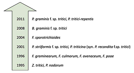
Development of polymerase chain reaction-based assays for the detection of wheat fungal pathogens from 1995 until the present. For each fungus, more detection systems were published later.
Development of polymerase chain reaction-based assays for the detection of wheat fungal pathogens from 1995 until the present. For each fungus, more detection systems were published later.
Fusarium graminearum. PCR RAPD and restriction fragment length polymorphism have been used first to study the genetic variation among strains of F. graminearum [Quellet and Seifert, 1993; Schilling et al., 1994]. F. graminearum detection systems by Schilling et al. [1996] and Nicholson et al. [1998] were based on SCARs. Schilling et al. [1996] showed that species-specific primers could not be designed for ITS1 and ITS2 regions of F. graminearum and F. culmorum due to the high sequence homology between these species. However, sufficient sequence variation was found in the ITS region to distinguish them from F. avenaceum. Nicholson et al. [1998] presented a PCR assay to detect and quantify F. graminearum in wheat. The Fg16F/R primer set produced a single DNA fragment of 400-500 bp and was successfully used in the quantitative analysis of F. graminearum in ear tissue of spring barley and winter wheat. Furthermore, Fg16F/R primers are used for molecular identification of other fungi belonging to FGSC. In some studies, it was observed that they amplified products of different size, i.e., about 400 bp in F. graminearum, about 550 bp in F. asiaticum and about 500 bp in F. meridionale [Castañares et al., 2014; Covarelli et al., 2011]. Jurado et al. [2005] developed species-specific primers for F. graminearum based on the IGS region. The Fgr-F/R primers were tested on the DNA obtained from wheat seeds with the confirmed presence of this species. gaoA gene is promising target for F. graminearum detection due to low homology with other fungi [Biazio et al., 2008]. GOFW/GORV primes targeting gaoA gene showed similar specificity that method describe by Schilling et al. [1996].
Fusarium culmorum. Similarly to F. graminearum, early F. culmorum detection was established based on the SCAR sequences [Nicholson et al., 1998; Schilling et al., 1996]. The Fc01F/R primers were able to identify all tested F. culmorum isolates, whereas the Fcg17F/R primer set was specific for both F. graminearum and F. culmorum [Nicholson et al., 1998]. Specificity of the Fc01F/R primer set only to F. culmorumspecies was further confirmed on environmental samples in the experiment conducted by Demeke et al. [2005]. This assay was also confirmed to be the most suitable for strains infecting cereals in Poland [Baturo-Cieśniewska and Suchorzyńska, 2011]. The detection rate of the assay with the OPT18F/R470 PCR primer set was estimated at 50 pg. It was specific for all 69 isolates tested [Schilling et al., 1996]. However, in a recent study they gave false negative results for 5 of 68 environmental isolates collected from different Polish provinces [Baturo-Cieśniewska and Suchorzyńska, 2011]. In the study of Sanoubar et al. [2015] primers Fc03/Fc02 amplified a 140 bp fragment of F. culmorum genome. They were easily adapted to quantitative analysis in order to investigate F. culmorum presence and deoxynivalenol production.
The methods of Mishra et al. [2003] and Jurado et al. [2005] targeted ITS and IGS of rDNA, respectively. The 175F and 430R primer set were tested in 92 isolates of 5 toxigenic Fusarium species and validated against the NCBI sequence database [Mishra et al., 2003]. Jurado et al. [2005] developed the Fcu-F and Fgc-R primer set tested on strains and environmental samples of wheat seeds with the confirmed presence of F. culmorum. In addition, Fcu-F and Fgc-R were later used for the analysis of fungi from various plant materials, e.g., maize [Covarelli et al., 2011].
Fusarium avenaceum.Schilling et al. [1996] and Turner et al. [1998] developed assays to detect Fusarium avenaceum species based on SCARs of the ITS region. The FA-ITSF and FA-ITSR primer set was tested on fungal isolates obtained from different habitats. No cross-reaction was observed for other Fusarium strains and plant species tested [Schilling et al., 1996]. Turner et al. [1998] developed Fa-U17F/R primers and reported that they were not only specific to F. avenaceum but also to F. tricinictum. Similarly as in previously mentioned SCAR-based assays, it indicated that the separation of Fusarium species could be problematic. Turner et al. [1998] also developed PCR primers (J1AF/R) that were verified in the study of Demeke et al. [2005]. The J1AF/R set was specific only for F. avenaceum isolates while the AF/R primer set revealed the incidence of F. avenaceumin the field plots in the study of Doohan et al. [1998]. These authors amplified all tested isolates of F. avenaceum ssp. avenaceum, but also F. acuminatum ssp. acuminatum [Demeke et al., 2005].
Fusarium poae.First and the most commonly used system for the detection of F. poae was based on RAPD assays [Parry and Nicholson, 1996]. The authors performed cloning, southern blotting of DNA from F. poae and other wheat seed and stem base pathogens. One fragment, which hybridized directly to F. poaeDNA was used to design the Fp82F/R primer pair. It amplified target DNA from all isolates of target species and gave negative results with any other fungal species associated with cereal diseases. Fp82F/R confirmed their specificity on the infected wheat seed samples. Konstantinova and Yli- Mattila [2004] described another primer set (CNL12/PoaeIGSr) that amplified a single 306-bp fragment from all F. poae strains. However, these primers cross-reacted with F. kyushuense and F. langsethiae. Similarly, Tox5-1/Tox5-poae R primers amplified DNA of all 45 isolates of F. poae and gave a non-specific product for one of F. langsethiaeisolates [Niessen et al., 2004]. Jurado et al. [2005] developed the Fps-F and Fpo-R primer set based on partial IGS sequences. These species-specific PCR primers amplified a DNA fragment only for F. poae isolates. Fps-F could anneal to DNA from any Fusarium species and showed specificity for particular species in pair with other primers [Jurado et al., 2006].
Fusarium sporotrichioides.In the Konstantinova and Yli-Mattila [2004] experiment, IGS-PCR products after enzymatic digestions generated specific restriction fragment length polymorphism fragments that were used for primer design. The CNL12/PulvIGSr primer set generated fragments of 610 and 750 bp in F. sporotrichioides and F. langsethiae, respectively, and cross-reacted with the plant genome but not with other pathogenic fungi. Konstantinova and Yli-Mattila [2004] developed also SporoITSf/SporoITSr primers that gave a signal for most F. sporotrichioides and F. langsethiae strains. However, these primers generated 2 unexpected bands for most strains of F. sporotrichioides and F. langsethiae and many nonspecific products for other species. Kulik et al. [2004] developed an assay to detect F. sporotrichioidesbased on ITS2. The FspITS2K and P28SL primer pair amplified a fragment of 288 bp. Of all of the primer sets in the experiment of Wilson et al. [2004], Fspo/LanspoR1 amplified a product of 332 bp from all of the tested isolates of F. sporotrichioides. It was developed based on the RAPD assay and required a small amount of template DNA, i.e., 100 fg. The 127F1/127F2 primer pair designed by the same authors was able to detect F. sporotrichioides and F. langsethiae.Wilson et al. [2004] also designed primers based on the Tri5 gene sequence. The Spo3F and Spo1R primer set was able to amplify a product of 641 bp with DNA of F. sporotrichioides, but also F. langsethiae.The Tox5-1 primer, as a universal forward primer together with a specific reverse primer (Tox5-sporo R2), gave a single 400-bp product with DNA isolated from most F. sporotrichioidesstrains [Niessen et al., 2004]. The AF330109CF/R primer set was tested on fungal cultures obtained from environmental field samples and all target isolates produced the expected 332-bp DNA fragment [Demeke et al., 2005]. Jurado et al. [2005] developed specific primers for F. sporotrichioides based on the IGS region. Fps-F and Fsp-R primers generated a fragment of 400 bp. Wolny-Koładka et al. [2015] used this primer set for the assessment of F. sporotrichioides prevalence in ears of winter wheat.
Puccinia graminisf. sp. tritici.Wang et al. [2011] developed the Pgtfssr1-F/R primer set that was able to detect P. graminisf. sp. tritici based on the microsatellite-enriched genomic library constructed using the method of FIASCO (fast isolation by AFLP of sequences containing repeats). The primers were tested with other fungal pathogens and infected wheat tissues. Chen et al. [2015] developed a multiplex PCR reaction for simultaneous detection of 3 pathogens, i.e., P. graminisf. sp. tritici, P. triticina, and B. graminisf. sp. tritici, with the use of the aforementioned primer sets. This strategy provided a rapid method for quick identification of several wheat pathogens in one tube. Liu et al. [2014], using the FIASCO technique, generated the Pgtw (f)/ Pgtw (r) primer set specific which id reliable for the identification of P. graminisf. sp. tritici. This method generated a 330-bp DNA fragment. No amplicons were obtained from other nontarget wheat fungal pathogens (P. triticina, P. striiformis, Tilletia controversa Kühn, T. caries, T. foetida, Ustilago tritici, Sporisorium reilianum, Gibberella zeae, and B. graminis f. sp. tritici). Berlin et al. [2012] used rust-specific primers (ITS1RustF10d and StdLSUR2a) from the study of Barnes and Szabo [2007] to identify P. graminis using both PCR and DNA sequencing. ITS1rustF10d and StdLSUR2a hybridized to ITS1 and 28s rDNA regions, respectively.
P. striiformisf. sp. tritici.Primer sets targeting the β-tubulin gene (YRNT1/YRNT2) were developed in the experiment of Fraaije et al. [2001]. PCR assay was tested against several other fungi and infected wheat leaves from different localities. In the study of Lihua et al. [2008], genomic DNA of different lines of P. striiformis and P. triticina was amplified using the BAF6/BAF2 primer set. Bands obtained with these primers were used to design 2 diagnostic SCAR primers, i.e., YR(f)/(r1) and YR(f)/YR(r2). They gave specific products of 160 and 139 bp, specific only for P. striiformis isolates. The universality of the latter primers was tested against 80 isolates of P. striiformis from different localities and several other pathogenic wheat fungi. No cross-amplification was observed. Zhao et al. [2007] designed the PSR/PSF primer set that gave a single product of 169 bp for only P. striiformisf. sp. triticiisolates with a maximum traceability of 0.1 pg of target DNA. The reliability of that assay was confirmed under greenhouse and field conditions with naturally infected leaves even beforeany visible symptoms. Wang et al. [2008] developed a specific and sensitive detection method based on the P. striiformis repeat (PSR) sequence suggested by Zheng et al. [2000a, b] as a useful target for specific detection by PCR. The specificity of PST2 f/r primers was tested and confirmed against healthy plant tissue, P. striiformis, and other 6 isolates of fungi. The analysis of wheat leaves inoculated with P. striiformis f. sp. tritici indicated that fungal DNA could be detected with this PCR assay in latent infected leaves (3 days after inoculation) and during early stages of infection. This primer set (PST2 f/r) was further examined in the experiment of Wang et al. [2009] as outer primers in nested-PCR with inner Nesta/Nests primers. Gao et al. [2016] developed 2 pairs of SCAR primers, i.e., PSTF117/PSTR363 and TF114/TR323. These primers produced 274- and 180-bp amplicons, respectively, which could detect the pathogen on wheat leaves even a few hours after inoculation.
P. triticina (syn. P. reconditaf. sp. tritici). Fraaije et al. [2001] developed multiplex PCR assay for simultaneous detection of P.recondita, Z. tritici, P. nodorum, and P. striiformis.The sequence of the β-tubulin gene was used as a target. The BR3/BR2 primer set gave a single 300-bp product with the traceability of 68 pg of fungal DNA. The second known PCR detection assay for P. triticina was proposed by Cao et al. [2007] and developed into multiplex assays by Chen et al. [2015]. The BAFBY primer set (f1)/(r) was successfully tested against Puccinia species, wheat leaves inoculated with P. graminisf. sp. tritici, P. triticina, and B. graminisf. sp. tritici, and noninoculated wheat leaves collected from the fields.
Zymoseptoria tritici.The first primers designed by Beck and Ligon [1995] were based on ITS regions. Amplification of fungal DNA with JB446 and ITS1 primers resulted in a 345-bp product. Fraaije et al. [1999] developed the E1/STSP2R primer set based on the β-tubulin gene of Z. triticiand compared their assay with Z. tritici-specific primers described by Beck and Ligon [1995]. E1/STSP2R primers allowed obtainment of a single product; however, PCR with JB446 and ITS1 resulted in a second product of about 280 bp, which was amplified from healthy plant tissue. Fraaije et al. [2001] developed another β-tubulin-based primer set (STIF2/BAF4ST) for Z. triticidetection. Similar to the ITS1/JB446 primer pair, both assays introduced by Fraaije et al. [1999, 2001] finally proved to give multiple bands from some other wheat pathogens [Guo et al., 2006]. Guo et al. [2006] proposed 4 newly designed primer pairs amplifying fragments of rDNA, the act1 gene, and RAPD fragment R5870-2. Primer pairs were tested with isolates of Z. tritici, other fungal pathogens, and healthy and inoculated plants. In the study of Consolo et al. [2009], primers were designed based on the alignments of ITS and α-tubulin sequences of the pathogen. Four sets of oligonucleotides amplified a single DNA fragment from all Z. triticiisolates and no product was obtained from other DNA sources.
Parastagonospora nodorum.Beck and Ligon [1995] developed JB433/JB434 primers for the detection of P. nodorumbased on ITS sequences. PCR assay demonstrated specificity, and no product was generated from Z. tritici-infected and healthy wheat tissues. Blixt et al. [2008] confirmed the usefulness of this assay for P. nodorum detection in wheat from Sweden. Specific primers (SNSP7F/CONS1R) were designed by Fraaije et al. [2001] in the sequence of the β-tubulin gene of P. nodorum. Malkus et al. [2005] amplified and sequenced full-length sequences of the tubulin gene (tubA). They developed primer sets from intron 1 and intron 3 sequences.
Blumeria graminisf. sp. tritici.Two assays for the detection of B. graminisf. sp. tritici were developed. Zeng et al. [2008] developed a primer pair able to specifically amplify B. graminisf. sp. triticiDNA. BF-F1/R PCR primers produced a single 464-bp product and were used by Chen et al. [2015] in the multiplex detection of 3 pathogens. Zeng et al. [2010] developed a nested PCR assay. The application of external and internal primer pairs improved its sensitivity.
Pyrenophora tritici-repentis.Mavragani et al. [2011] developed Pyr1/2 primers that were able to detect Pyrenophora and Drechslera species. Pyr1/2 were used as internal primers in nested PCR together with ITS1-F and ITS4 as external primers. Primers amplified a 344-bp product with P. tritici-repentis DNA as a template. Hudcovicova et al. [2015] described another PCR assay specific for P.tritici-repentis. The DTR1-F/R primer set amplified a 382-bp amplicon from ITS1-ITS2 regions of rDNA. New PCR assay did not cross-react with DNA from other wheat pathogens and healthy plant tissue.
Future Perspectives
Detection and identification of plant pathogens are essential for successful disease management. Conventional detection methods based on disease symptoms or laboratory identification by morphological, cultural, and rarely biochemical criteria can only be employed by qualified personnel with extensive knowledge in taxonomy. These time-consuming methods do not always provide unequivocal results, as they do not allow separation of closely related species. PCR diagnostic assays do not have most of these disadvantages, and they allow detection of all of the relevant wheat pathogenic fungi [Fones and Gurr, 2015; Fraaije et al., 2001; Keon et al., 2007; Shetty et al., 2007; Zeng, 2010].
McCartney et al. [2003] described PCR applications in plant pathology. These areas include plant disease control improving decision making regarding the use of fungicides and resistant cultivars, the detection of mating and virulence types, monitoring crop exposure to pathogen inoculum, and assessment of the pathogen population structure. One of the biggest advantages of PCR-based methods is the diagnosis of a presymptomatic disease presence [Keon et al., 2007; Shetty et al., 2007].
This review describes a number of PCR tests developed during the last 2 decades (1995-2015). This article points out that there are many PCR assays known for certain fungi species occurring on wheat, whereas for other crops the number of detection systems is very limited. There are many detection systems for F. sporotrichioides, P. striiformisf. sp. tritici, and Z. tritici [Beck and Ligon, 1995; Consolo et al., 2009; Demeke et al., 2005; Fraaije et al., 1999, 2001; Gao et al., 2016; Guo et al., 2006; Konstantinova and Yli-Mattila, 2004; Lihua et al., 2008; Niessen et al., 2004; Wang et al., 2008, 2009; Wilson et al., 2004; Zhao et al., 2007]. Particularly for Z. tritici, the number of known diagnostic tests is justified by the significance of STB, one of the most important foliar diseases of wheat caused by this fungus [Keon et al., 2007; Shetty et al., 2007]. A very limited number of conventional PCR protocols is known for F. graminearum, P. graminisf. sp. tritici, P. triticina (syn. P. recondita f. sp. tritici), and P. tritici-repentis [Cao et al., 2007; Fraaije et al., 2001; Hudcovicova et al., 2015; Jurado et al., 2005; Liu et al., 2014; Mavragani et al., 2011; Nicholson et al., 1998; Schilling et al., 1996; Wang et al., 2011]. These pathogens also cause wheat diseases of great economic importance; therefore, new PCR assays detecting these fungi should be developed. Another rationale for the development of PCR systems is a phenomenon of a fungus latency period, when visual inspection cannot determine the presence of a fungus, but DNA-based methods could be useful for pathogen detection [Fones and Gurr, 2015; Keon et al., 2007; Shetty et al., 2007; Zeng, 2010].
In recent years, several new molecular techniques have been proposed for pathogen detection. Quantitative PCR (qPCR, real-time PCR) has become one of the most promising techniques for the detection and characterization of phytopathogenic fungi. The sensitivity of qPCR is similar to or better than that of other PCR methods; however, it should be underlined that quantification of trace amounts of fungal DNA is often challenging. This is mainly caused by the relatively low fungal load in environmental samples and the structure of the fungal cell wall, which makes its disruption for nucleic acid extraction difficult [Leite et al., 2012]. For example, the detection limit for DNA extracted from M. graminicolacultures was 100 pg/μL using conventional PCR, whereas it was only 50 fg/μL by qPCR; thus qPCR assay was 20-fold more sensitive than conventional PCR [Guo et al., 2006]. PCR modification involving probe hybridization with fluorescent dye is known as fluorescent amplification-based specific hybridization (FLASH-PCR). In contrast to qPCR, the detection of fluorescence does not occur during amplification but rather after its termination. It also does not require specific equipment or electrophoresis. This method was tested in the identification procedures for P. nodorumand Z. tritici as well as toxigenic Fusarium species [Abramova et al., 2008; Ryazantsev et al., 2008]. Loop-mediated isothermal amplification, as opposed to PCR, does not require highly purified DNA or specialized equipment, because this method is performed at a constant temperature and no postamplification manipulations are necessary. This enables performing molecular detection on site. According to Niessen [2013], loop-mediated isothermal amplification assay has a high potential for future plant pathology studies. The detection of several DNA sequences at the same time was also achieved with the use of next-generation sequencing [Ronaghi, 2001]. However, bioinformatic data analysis was problematic for environmental samples because of the numerous nucleic acids of different origin in samples that can serve as potential sequencing templates, yet, this metagenomic approach was adjusted to detect phytopathogens by introducing unique pathogen-specific sequences, i.e., e-probe avoidance, e.g., data generated from the host plant [Stobbe et al., 2013].
Molecular diagnostics using conventional PCR systems seems to be the most cost-effective option compared to other alternative molecular methods. PCR-based diagnostics has a huge impact on mycology development but also in monitoring of good agricultural practice and good manufacturing practice. Proper identification of wheat pathogens is not without significance for the health of plants, animals, and humans. However, there is a limited number of PCR assays for certain species, which in turn indicates that further work in this area appears to be necessary.
Acknowledgments
This article was written with financial support from the National Centre for Research and Development, V LIDER Programme (LIDER/014/263/L-5/13/NCBR/2014), Poland.
Disclosure Statement
The authors have no conflicts of interests to declare.
